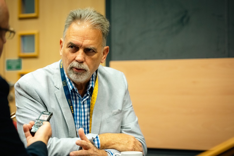
Stem cells implanted near spinal cord injuries spring into action to produce anti-inflammatory proteins – a function they do not display when isolated. This remarkable form of adaptation is what guided Prof. Dr. Antal Nógrádi’s research group in identifying the proteins most suitable for treating experimental spinal cord injuries in rodent models. To deliver the proteins, they began utilizing mRNA-based therapy as early as 2019, before the COVID-19 pandemic. Their cutting-edge research on mRNA treatments also garnered significant attention at the mRNA Conference hosted by the University of Szeged and funded by the Novo Nordisk Foundation. Professor Nógrádi, Head of the Department of Anatomy, Histology, and Embryology at the University of Szeged’s Albert Szent-Györgyi Medical School, gave an interview following his presentation at the conference.
Q: Professor, your group has made significant strides in spinal cord injury research through stem cell implantation in recent years. For example, using a rodent model, you were able to clarify the mechanism of action of proteins produced by stem cells. Given that stem cell treatment had been a successful research direction for you, why did you shift to working with mRNA therapy?
A: In the 2000s, we began focusing on salvaging damaged motor neurons, as there are conditions where these motor nerve cells are destroyed by spinal cord diseases or injuries but can still be preserved if intervention occurs during the acute phase. One of our initiatives was to launch stem cell therapy experiments in 2007, aided by Professor Ernő Duda, who had a high-quality mouse stem cell line preserved in his freezer, originally isolated by him and Professor Emília Madarász. Unfortunately, a substantial portion of stem cells are unsuitable for implantation. However, we found that our cell line could, in fact, be used to salvage damaged nerve cells, so we set out to understand how it functions. At the time, there were many proponents of the idea that implanted stem cells could differentiate and transform, effectively substituting the destroyed cells and enabling cell replacement. Yet, we observed only minimal evidence of this, being able to identify just a few new myelin-forming cells derived from this cell line. What we did detect was a so-called humoral mechanism – associated with molecules produced by cells – in which the stem cells, when implanted near the injury site, generated what is known as a ‘secretome’ consisting of four to five factors. We named this secretome ‘injury-induced secretome.’ Once we identified its composition, we decided to move away from the entire stem cell implantation approach – as we were aware that no one would want to be injected with mouse stem cells or their modified variants. Instead, our aim was to learn from the humoral mechanism observed in the stem cells and build our research upon that foundation. In the subsequent phase, we found this approach to be effective, and we were successful in replicating the results simply by independently introducing the produced secretome, without the need for stem cells. However, this presented a rather complex challenge, leading us to consider using mRNA technology. Our plan was to have the modified mRNA that codes for the secretome factors delivered directly to the injury site. This idea prompted us to reach out to Norbert Pardi at the University of Pennsylvania in 2019, knowing he had been using mRNA technology based on the research done by Katalin Karikó and Drew Weissman.
Q: Prior to the pandemic, mRNA-based therapy was far from the focus of scientific attention. What did you and your team know about it at the time?
A: Back then, none of us were familiar with the intricacies of mRNA therapy for the nervous system. We did understand what mRNA was and knew that research into its therapeutic applications was underway, but lacked insight into how it would function in the context of spinal cord injuries. While naked mRNA may work when forced into a cell, its delivery poses significant challenges. Although we believed it could still function in a cell culture, we were also aware that the dynamics change dramatically in in vivo experiments involving living animals. However, by the time we reached out to Norbert Pardi, lipid nanoparticles for mRNA delivery had already proven to be highly effective. Here, I should note that it took Dr. Pardi a year and a half to convince the Canadian biotechnology company Acuitas of the effectiveness of lipid encapsulation – proving that progress doesn’t happen overnight there, either. With the initial mRNA package, we began by exploring how a simple reporter protein, GFP, which induces green fluorescence, would reach its intended target. We found that the cells absorbed and then expressed the protein coding for the mRNA, and we confirmed that this occurred in both intact and injured animals. We then progressed to introducing a therapeutic protein, IL-10, which was one of the factors we identified in our secretome. Due to the fact that IL-10 is an anti-inflammatory protein that has an effect on its own, we decided to explore the impact of IL-10 mRNA through separate experiments. Given that neurons carry the IL-10 protein receptor, we anticipated that the complete protein could bind to nerve cells. By analyzing the results from treated animals, we found that 60 percent of the cumulative effect of the four proteins identified in the secretome was attributable to IL-10. While this confirmed IL-10’s neuroprotective and anti-inflammatory properties, we still needed a protein to promote the growth of axons (or extensions) of surviving and damaged nerve cells. That protein was GDNF, which was also present in our secretome.

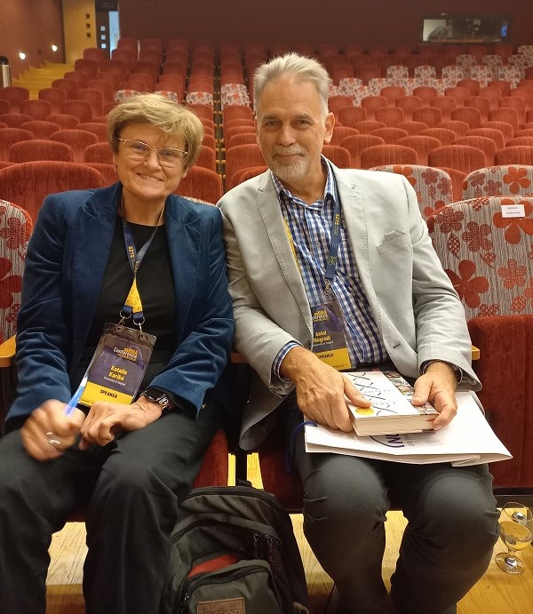
Katalin Karikó and Antal Nógrádi during a break at the mRNA Conference
Q: It’s intriguing how stem cells can so precisely secrete beneficial substances to address an injury in another organism! Have you ever considered why that might be?
A: While we may not be able to precisely articulate the reasons, it is evident that the stem cells fulfilled their role in an intelligent manner, successfully assembling an effective secretome in the vicinity of the injury. Consider this: unlike many other cases, this isn’t a situation where stem cells produce a secretome in their baseline state, carry it to the injury site unchanged, and refuse to adapt. On the contrary, these stem cells, in their native state in a cell culture, do not produce substances beneficial to us. However, once introduced into an injured environment, they adapt to the given injury type and, through various interactions, begin producing a secretome composition optimized for that specific injury. This process continues until the stem cells begin to transform and differentiate, at which point protein production ceases. I should add that this posed no issue for our research, as what we actually needed was the therapeutic window provided by the proteins in the secretome.
Q: As a layperson attending your conference talk, I found it fascinating to learn that there is a second wave of inflammation that occurs in spinal cord injuries.
A: In fact, there are multiple waves of inflammation! Initially, a strong first wave occurs due to the injury, bringing in macrophage scavenger cells that clear away foreign elements. Days later, a second wave begins, triggering a different inflammatory mechanism. Although macrophage cells arrive during this phase as well, our primary observation is that the expansion of the cavity resulting from the spinal cord injury arises from the deterioration of the damaged cells. At this stage, these deteriorating cells may still be salvageable. While the physical damage caused by the injury cannot be reversed, it is possible to intervene before the second wave of inflammation to prevent further cavity expansion. As for the subsequent waves, it has been documented that, surprisingly, additional waves of macrophages arrive in the spinal cord even weeks later, despite the injury already transitioning into a subacute and later chronic phase. The significance of these later waves remains unclear; however, we hypothesize that their actual role is to clear the area as inflammation subsides and a fixed, scarred state is established.
Q: So, if I understood correctly, the time window is between the first two waves?
A: Yes, the window opens between the two waves, around day 6 or 7. By this time, the first wave of macrophages has already subsided. Peak infiltration actually occurs on the fourth day and then begins to decline. There’s a critical point when the cavity starts to clear, the second phase has not yet begun, and the first wave is tapering off – this is when a window for cell therapy can be effectively utilized. Of course, there is also an immediate treatment opportunity right at the onset of the injury. If an effective molecule is introduced externally at that stage, it can penetrate, as the blood-brain barrier is already compromised and permeable.
Q: Should we envision the process as injecting mRNA encapsulated in lipid nanoparticles directly into the injured spinal cord?
A: That was our initial approach. Modified mRNA packaged in LNPs can indeed be injected. It is important to note that RNA itself is a very small molecule, but once it’s packaged, its size increases significantly, which poses a quantitative limitation. I also mentioned that by the seventh day, a cavity begins to form in the spinal cord at the site of the injury. This contusion cavity is tiny, with a volume of just a few microliters, making it smaller than a droplet. So, only a limited amount of material can be introduced into it without causing further damage or expanding it. However, exposing the injured spinal cord and injecting something into it represents an additional trauma, which is far from ideal at the end of the primary inflammatory phase; and when we wanted to deliver all four factors of the secretome into this closed space, it became clear that we were pushing the limits. So, we began exploring alternative pathways. Interestingly, the fact that certain parts of the body have macrophage reserves is nearly undocumented in the literature. Yet, adhered to the peritoneum, tiny cells lie dormant, waiting to mobilize toward an inflammatory site to clean up there. Our initial findings actually indicate that introducing mRNA into this confined space leads to two possible outcomes: the mRNA either enters systemic circulation and heads toward the liver, or it is promptly taken up by macrophages, which convert the molecule into a substantial amount of protein within hours – as our observations indicate. Simultaneously, the cells receive signals directing them to the spinal cord injury site, where the problem manifests itself, and they transport the therapeutic proteins there. Indeed, this method of application shows potential for use in humans, too.
Q: The insights you shared about this therapeutic application at the mRNA conference were met with great enthusiasm. Are there any other similar ideas your team is currently exploring?
A: In routine practice, such as with young children, the last-resort method for fluid replacement involves using the so-called intraosseous route, i.e., the vascular network of the bone marrow. This technique, commonly used in emergency care and even by paramedics, involves drilling a small hole into the tibia and delivering fluids through a cannula inserted into the bone, using commercially available instruments. The tibia contains a rich network of capillaries, which immediately absorbs the fluid. This network has the added advantage of allowing red blood cells and other formed elements of the blood from the marrow to be washed into circulation, thereby not only replenishing the body with the fluids but also compensating for blood loss. While this pathway could also be utilized, we still lack sufficient knowledge about what happens when mRNA is introduced into the bone marrow. Indeed, this is the research direction we plan to explore in the near future at the Biological Research Center in Szeged in collaboration with Zsolt Zimmermann and Miklós Erdélyi.

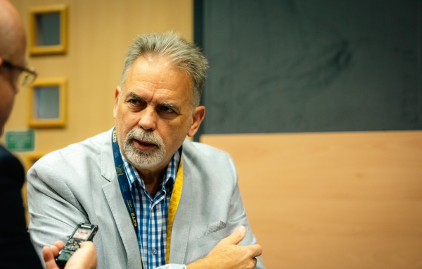
Prof. Dr. Antal Nógrádi, Head of the Department of Anatomy, Histology, and Embryology at the Albert Szent-Györgyi Medical School of the University of Szeged
Photo: Ádám Kovács-Jerney
Q: Have you observed any side effects associated with the delivery of mRNA?
A: When we began exploring mRNA therapy, our first step was to monitor any potential inflammation or side effects following the administration of mRNA to a healthy, intact animal. This approach was necessary because it’s challenging to monitor newly induced inflammation in an inherently inflamed environment. By then, it was known that the lipid nanoparticles encapsulating the mRNA could cause a very mild inflammation. In fact, it is necessary for them to induce some degree of inflammation to attract the macrophages, which are essential for making mRNA vaccination effective. In our experiments, however, we observed no measurable changes in the spinal cord. This was a highly positive outcome, as it indicated that LNP does not disrupt the immune system of a healthy animal. That said, it’s important to note that the situation in the spinal cord differs from that in the peripheral immune system. Even after a minor puncture, the spinal cord maintains its protected immunological state. In professional terminology, this is referred to as a privileged immune state – which is indicative of the fact that immunological processes in the central nervous system function differently from those in the peripheral immune system. This protective mechanism is an evolutionary heritage, ensuring that the functioning of the central nervous system is tightly safeguarded by a number of mechanisms, such as the blood-brain barrier and immunological barriers.
Q: Speaking of evolution: if a stem cell is capable of producing “repair” proteins, why doesn’t the body itself develop new nerve cells?
A: Despite its efforts, it cannot succeed. That’s because the central nervous system of mammals is a developmentally finalized system, with the division of nerve cells being completed in the early embryonic stages. The number of neurons we have becomes fixed before birth and will not increase – the number is final; and there are several reasons for this. However, in animals that continue to grow throughout their lives, such as fish, the nervous system also grows continuously. The nerve cells of these animals keep dividing throughout their entire lifespan, which endows these creatures with immense regenerative potential. This shows that the nervous system itself is not incapable of regeneration; however, its growth is constrained by the rigid, non-expanding structures of the skull and spinal canal. As a result, when an injury occurs, the body’s response is to quickly repair and clean up everything. While regenerative processes do begin, inhibitory mechanisms are also activated. These mechanisms suppress excessive regeneration almost immediately. We have observed, however, that macrophages entering the spinal cord without any treatment actually produce IL-10 to a limited extent. Additionally, the body’s own neurons begin generating small amounts of IL-10. Yet, this quantity is insignificant compared to therapeutic levels – it doesn’t even qualify as a suboptimal dose. Even if it may contribute to stabilizing the situation, it falls short of providing a solution. That is why it is fortunate that in our experiments the stem cells “knew” what to do and thereby provided us with valuable insights into the factors that could be beneficial in this context.
Q: What will your next step be after achieving such results in the rodent model? What model do you plan to use next?
A: I believe it is essential to evaluate the treatment system in a larger animal model. So, we are actively seeking collaborations with professional groups – particularly those affiliated with veterinary universities – that are working on innovative treatments for domestic animals, especially dogs. Certain dog breeds, such as dachshunds, are particularly prone to spinal diseases due to the so-called “long vehicle” principle, which relates to the spine’s role as a supporting structure. Consequently, the spine is unable to sustain its excessively elongated load-bearing arch, ultimately leading to its collapse. One treatment approach involves stabilizing the spine as a physical, bony structure; another focuses on treating the affected spinal cord itself. This provides an excellent opportunity to work with a high-quality animal model and a genetically uniform dataset, with the consent of the pet owner. Moreover, direct intervention in the spinal cord may not even be necessary, as our innovative methods open up the possibility of treating these animals by stimulating IL-10 production in macrophages originating from the abdominal cavity.
Original Hungarian text by Sándor Panek
Photos by Ádám Kovács-Jerney
In the feature photo: Prof. Dr. Antal Nógrádi, Head of the Department of Anatomy, Histology, and Embryology at the Albert Szent-Györgyi Medical School, University of Szeged

