Case 5.: Case of a young female patient carrying congenital R24P CDKN2A gene mutation with multiple primary tumors
In December 2006, a 1.524 mm thick, pT2b, Cl. II-III melanoma malignum has been removed from the left waist region of a 31-year-old female patient. From the left inguinal region, 1 sentinel lymph node, from the left axillary region, 4 sentinel lymph nodes have been excised with a negative histological result. As a postoperative treatment, she received interferon therapy between March 23, 2007 and February 7, 2008 at the SZTE Department of Dermatology.
On February 14, 2008, the patient noticed a walnut-sized lump above the left pectoral major muscle that has been removed at the SZTE Department of Dermatology with the following histological result: lymph node metastasis of anaplastic breast cancer, ER,PR: negative, HER-2: negative.
In further search of a primary tumor the following examinations were done:
Abdominopelvic CT (April 9, 2008): The head of the pancreas was 4-5 mm thicker.
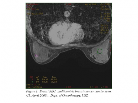
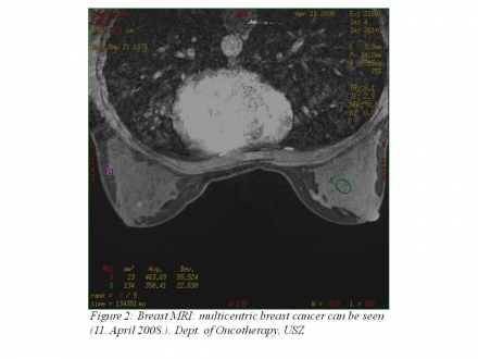
Breast MRI (April 11, 2008): on the left side in the breast tissue several 3-8 mm contrast-enhancing lesions can be seen (Figure 1 | Figure 2). A 13 mm lymph node in the axilla.
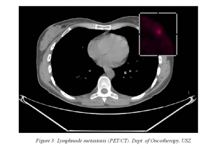
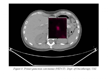
PET CT (April 30, 2008): A 1 cm FDG accumulation at the border of the upper quadrants in the left breast. In the left axilla, a 7.16 mm accumulating lymph node is visible (Figure 3). At the border of the body and the tail of the pancreas, a 1.5 cm accumulation can be seen (Figure 4).
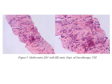
In possession of the breast MRI and the PET/CT results, based on the decision of the oncology team, mastectomy and ABD was performed on May 13, 2008 at the SZTE Department of Surgery. The histological result of the mastectomy was in concordance with the result of the excision performed in February 2008: 4 foci of IDC (Figure 5), Grade III. (3+3+3) pT1 (12 mm), vascular invasion, pN1 (2/14), extracapsular invasion, closest margin of resection 8 mm, ER: negative, PR: 60%, HER-2: negative.
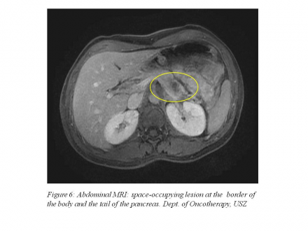
Regarding that PET/CT described a 15 mm accumulation at the border of the body and the tail of the pancreas the following imaging examinations were performed: Abdominal MRI (June 13, 2008): a 2 cm space-occupying lesion can be seen at the border of the body and the tail of the pancreas (Figure 6). ERCP (June 26, 2008): a stop can be detected at the border of the body and the tail of the pancreas.
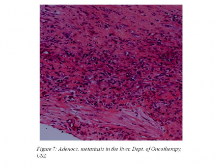
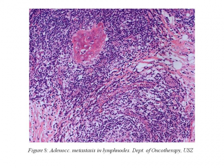
According to the decision of the oncology team, on July 1, 2008 explorative laparotomy, liver biopsy and lymph node biopsy were performed. Histological description revealed an adenocarcinoma with a liver (Figure 7) and lymph node (Figure 8) metastasis.
As an adjuvant treatment of her breast carcinoma and a palliative treatment of her metastatic pancreatic carcinoma, the 33-year-old female patient received 6 cycles of gemcitabine-cisplatin chemotherapy between July 18, 2008 and January 15, 2009 at the SZTE Department of Oncology.
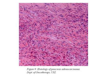
Before the start of chemotherapy, only CA 19-9 (152.4) was elevated among her tumor markers. During chemotherapy, control chest and abdominopelvic CT examinations and the control of tumor marker levels were performed 3 monthly. Control abdominopelvic CT examinations showed continuous regression at both 3 months and at 6 months, while chest CT and tumor marker results did not show any abnormalities. As side effects of the chemotherapy, Grade I nausea, vomiting, diarrhoea, Grade II alopecia, anaemia and neutropenia developed. Because of Grade II anaemia and neutropenia the patient received GCSF (Granulocyte Colony Stimulating Factor) and EPO (erythropoietin) treatment via subcutaneous injections. Cycle postponement occured twice during chemotherapy, due to side effects. An abdominopelvic CT made on January 27, 2009 showed complete remission of the pancreatic process. A PET CT made on March 3, 2009 also brought a negative result, so according to the decision of the oncology team pancreatic tail resection and splenectomy was performed on May 19, 2009 at the SZTE Department of Surgery. The histological examination did not reveal any malignant tissue, a picture of chronic fibrotizing pancreatitis (Figure 9) could be seen in the pancreas. The patient was tumor-free until December 2011 and her tumor markers are within the normal range. At that time, two new primary malignant melanomas were removed from the skin of the abdomen.
In the family history of the young female patient 2 close relatives were also diagnosed with tumor. Her father had laryngeal and gastric tumor, he is tumor-free after an adjuvant treatment. Her paternal aunt died of breast cancer at a young age. Genetic examination was performed at the patient and her parents. BRCA1 and BRCA2 mutations were not detected at any of the relatives, but it turned out that both our young patient and her father carry R24P CDKN2A gene mutations. This gene mutation predisposes to the development of synchronous and metachronous tumors, especially melanoma and pancreatic tumor.


