Case 2.: Case of a fulminant breast cancer
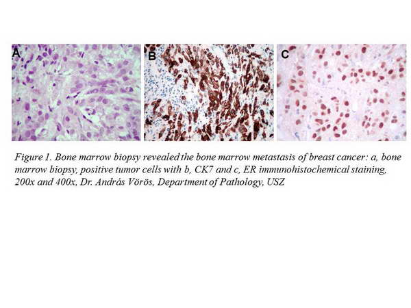
The 59-year-old female patient, who had a negative breast screening examination 10 months earlier, had been investigated because of anaemia and pancytopenia. Abdominal ultrasound, gastroscopy and chest x-ray did not show any significant abnormalities, so a bone marrow biopsy was performed. Pathologic examination revealed a bone marrow metastasis of a desmoplastic carcinoma that turned out to be estrogen receptor positive (Figure 1).

In the light of the pathologic diagnosis breast or endometrial cancer could be supposed as a primary tumor. As a search for primary tumor and metastases the following examinations were performed with the results below.
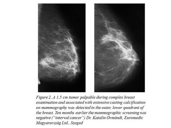
On chest CT there was no sign of primary lung tumor or metastasis, but bone scan did not rule out the possibility of metastasis in case of the nodules seen in the projection of the ribs. Tumor marker levels indicated a malignant process: CA 15-3: 2355, CA 125: 116.09, CEA: 4.4, CA 19-9: 2.73. During a complex breast examination a palpable, 1.5 cm lump had been found in the outer, lower quadrant of the left breast (Figure 2).

This lump turned out to have a radiologically malignant morphology, and an ultrasound guided biopsy had been taken from it. No abnormality was found in the opposite breast and the lymph node regions. The therapy-resistant disease with a fulminant progression caused the death of the patient a few months after the first symptom.
Edited by Prof. Dr. Zsuzsanna Kahán


