SZTE Nanomedicine Research Group
Principal investigator: Prof. Dr. Ildikó Csóka
Preparation and characterization of liposomal nanostructures for intranasal drug delivery

Liposomes are nanosized lipoidal vesicles that are under extensive investigation as drug carriers with improving the delivery of therapeutic agents. Liposomes can trap both hydrophobic and hydrophilic compounds. The active agent is needed to be protected or when the solubility is poor, this time we can use the liposomes in the intranasal intake.
The liposomes have a lot of advantages. They increase the efficiency of pharmacones and reduce side effects.
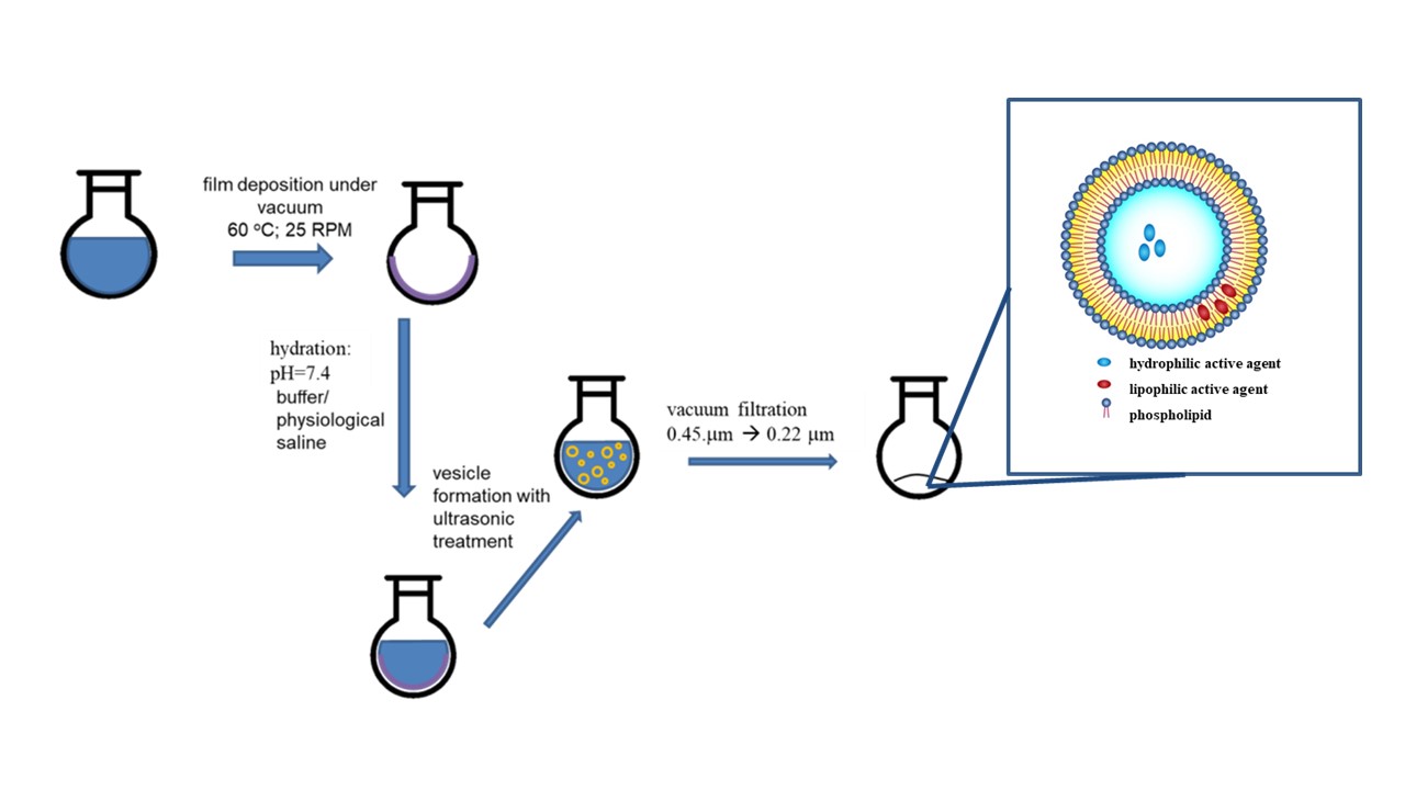
The method of Bangham et al. is one of the simplest procedures for liposome formation. Here, liposomes were prepared by first hydrating a thin lipid film in an organic solvent (ethanol or chloroform-methanol mixture), which is then removed in a subsequent film deposition under vacuum. The solid lipid mixture is then hydrated using an aqueous buffer or physiological saline solution. The lipids spontaneously swell and hydrate to form the liposome. In the last step ultrasonication and membrane vacuum filtration is applied. The liposomes were stabilized via lyophilisation ensuring their long-term stability.
The liposome’s properties are determined by various analytical techniques. The morphology of the samples examines by transmission electron microscopy (TEM). Dispersion of the sample particles is drop-casted onto a 200-mesh carbon-coated copper TEM grid and dried in air. TEM images are obtained using a FEI Tecnai G2 20 Twin HR-TEM electron microscope operated at 200 kV.
Thermogravimetric analysis (Mettler Toledo TGA/DSC1 STARe System) use to investigate the decomposition of the as-prepared and lyophilized liposomes. The particle size distribution determines by laser light scattering on a Particle Size Analyzer (Zetasizer Nano ZS, Malvern Instrument), while the mean vesicle size, the polydispersity index (PDI) and the surface charge of the prepared samples are also determined.
The weight of the dried nanospheres for the TGA experiment is between 5 and 10 mg. The heating curve record under 20 ml/min nitrogen flow at a 10 °C/min scan rate between 25 and 600 °C. The glass transition temperature (Tg) of the liposomes determine by differential scanning calorimetry (Mettler Toledo DCS821e). The IR active covalent functional groups characterization by Fourier-transform infrared spectroscopy (Avatar 330 FT-IR Thermo Nicolet) in KBr pellets in wavenumber range from 2000 to 700 1/cm.

